Introduction
Traumatic dental injuries, particularly fractures of the anterior teeth, are a significant concern in dental practice due to their impact on both function and aesthetics.1 In cases involving the upper incisors, patients often experience psychological distress in addition to the physical consequences of the injury, as damaged front teeth can affect appearance, speech, and self-confidence. Tooth reattachment offers a minimally invasive and highly aesthetic solution by preserving the natural tooth structure and restoring the original anatomy.2 This technique is particularly beneficial when a fractured tooth fragment is available and intact, allowing for a superior color match and texture compared to other restorative materials.
The success of tooth reattachment relies on various clinical factors, including the site and size of the fracture, pulpal involvement, and periodontal health. With the advancement of adhesive dentistry and reinforcement techniques, such as the use of fiber posts and ribbond fiber, the structural integrity of reattached teeth has improved significantly. These materials distribute stress more evenly and enhance the longevity of the restoration, mimicking the biomechanical properties of natural teeth.3
This case report presents the management of an Ellis Class III fracture in a 32-year-old female patient, utilizing fiber reinforcement and modern adhesive techniques to achieve both functional and aesthetic restoration.
Case Presentation
A 32-year-old female patient presented with the chief complaint of a broken upper front tooth (Figure 1). She reported a history of a fall a few hours prior, which was associated with severe pain. Clinical examination revealed an Ellis Class III fracture involving the enamel, dentin, and pulp tissue of tooth #22, with associated pain and tenderness on percussion. While there was no intraoral swelling, extraoral soft tissue swelling was noted on the lower lip and mandible. The fractured tooth segment was carefully removed and stored in cold milk to preserve it for the reattachment procedure.
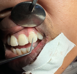
Figure 2
Carefully removed fractured segment atraumatically ,pulp tissue removed and disinfected, kept in cold milk until completion of re attachment procedure


Figure 5
Pulp tissue removed from chamber & disinfected, Tooth preparation done with 37%phosphoric acid etching and bonding agent applied.


Local anesthesia (lignocaine adrenaline injection-LOX 2% adrenaline 1:200,000) was administered using an infiltration technique around the fractured tooth to ensure effective pain relief and facilitate treatment. All procedures were performed under rubber dam isolation. The fractured segment was removed atraumatically, the pulp tissue was extracted from the chamber, and the area was disinfected (Figure 2).
The treatment plan included a single-visit root canal treatment with sectional obturation using gutta-percha and AH Plus sealer to address the pulpal involvement and alleviate the patient's pain (Figure 3). This was followed by a gingivectomy and osteotomy (Figure 2) to manage the subgingival fracture and restore proper biologic width.
When a fracture invades the biological width of a tooth, it is essential to restore both the biological integrity and function of the tooth while preserving as much of the supporting bone structure as possible. A surgical procedure involving minimal osteotomy and osteoplasty was performed in this case. The fractured tooth segment and the remaining portion of the root were meticulously prepared by etching (Figure 4) and applying a bonding agent (Tetric N Bond) based on the total etch technique. A glass fiber post (3M™ RelyX™ Fiber Post 3D Glass Fiber Post) was employed for post and core reconstruction, offering substantial reinforcement to the tooth structure. Coronal reinforcement was achieved using ribbond fiber, which has multi-directionally oriented fibers strategically wrapped to enhance stability and distribute stress effectively (Figure 6).
Bottom of Form


Discussion
The success of a tooth reattachment procedure depends on several key factors. The site of the fracture is crucial, as fractures near the gingival margin or involving the root can complicate the procedure, whereas coronal fractures are generally easier to manage.4 The size of the fractured fragment significantly affects stability, with larger fragments providing better bonding surfaces. Smaller fragments may lack sufficient support. Healthy periodontal tissues are crucial for healing and long-term success, while periodontal disease can compromise the prognosis. Other key factors include pulpal involvement, root maturation, biological width invasion, occlusion, and the timing of reattachment, as prompt intervention is vital for successful bonding and healing. All these factors must be carefully considered for optimal patient outcomes. 5
Tooth fractures are categorized as uncomplicated or complicated. Uncomplicated fractures involve only the enamel and dentin and are easier to manage since they don't affect the pulp. Complicated fractures involving enamel, dentin, and pulp are more challenging. Though less common, complicated fractures are harder to treat as they often lead to pulp inflammation or contamination.6 Tooth reattachment offers key advantages, primarily in esthetics, as the natural tooth fragment provides a perfect color and texture match. It also reduces tooth wear by preserving the original structure. The procedure is quicker, needing fewer appointments and less chair time compared to crowns or veneers. Additionally, using the original tooth leads to more predictable long-term results, ensuring a stable, natural outcome when done correctly.7 Supragingival fractures allow straightforward reattachment, but subgingival or intraosseous fractures need complex methods. Orthodontic extrusion or surgical techniques like crown lengthening, electrosurgery, or flap elevation expose the fracture. A minimal osteotomy or osteoplasty can restore integrity while preserving bone.
In this case report, the fractured tooth fragment was in sound condition and exhibited an excellent fit over the radicular portion of the tooth. Given this favorable scenario, reattachment of the fragment was deemed the most appropriate treatment option. To enhance the stability and longevity of the restoration, the use of a fiber post was ideal and provided additional support to the reattached fragment, reinforcing the structural integrity of the tooth and ensuring better bonding. This approach not only preserved the natural tooth structure but also offered a more durable and aesthetic outcome. When combined with a composite core and advances in adhesive techniques and materials, fiber posts enable the creation of a monoblock—a multilayered structure without weak interlayer interfaces. This approach reinforces the tooth by creating a unified structure, which mimics the strength and integrity of a healthy, unfractured tooth. The monoblock concept significantly enhances the overall stability of the tooth and helps restore its original biomechanical properties. Additionally, the use of Ribbond, a ribbon-like reinforcement material, further distributes stress across the remaining radicular dentin. This added reinforcement helps to protect the tooth from future fractures and enhances the longevity of the restoration.8 To support the repair and ensure optimal healing, the tooth was splinted with ribbond fiber for a duration of two weeks.
Resin-based sealers are recommended for obturating teeth planned for restoration with glass fiber posts, as eugenol-based sealers may inhibit the setting of resin cements.
Luting the fiber posts with resin cement not only reinforces the tooth but also helps achieve higher bond strengths of the fractured segments. It also minimizes the inclusion of air voids and is easy to use, making the procedure predictable. Dual-curing systems are the most suitable material, as they allow polymerization even in areas that are difficult for light to reach.9 Studies have shown that reattached tooth fragments can have success rates as high as 90% when evaluated based on parameters such as periodontal health, pulpal status, and color harmony over a follow-up period of up to 24 months. This high success rate indicates that, with proper care and technique, reattachment can be a reliable and long-lasting restorative option.10
Reattachment procedures not only aim to restore the stability and function of the tooth but also address the psychological aspect, especially in young patients who are often eager to retain their natural teeth. In this case, the patient, being young, expressed a strong desire to keep her tooth. She was fully informed about the treatment plan, its temporary nature, and the prognosis. In cases of fracture, reattachment should always be considered, as it is a more conservative approach compared to conventional post-and-core techniques or prosthetic rehabilitation, preserving as much of the natural tooth structure as possible. In this case, the outcome of the reattachment procedure was comparable to that of a crown. However, it is essential to understand that each case is unique, and the final treatment decision should be tailored to the specific situation and the patient's preferences. Options such as reattachment, prosthetic rehabilitation, or immediate implants should be carefully considered, taking into account the individual clinical circumstances and the patient’s desires.
Conclusion
Reattachment procedures are a valuable option for restoring tooth stability and function while addressing the psychological needs of patients, particularly younger individuals who are keen to retain their natural teeth. This conservative approach preserves more of the natural tooth structure compared to conventional post-and-core techniques followed by prosthetic rehabilitation. While the results of reattachment can be comparable to more invasive treatments, the decision should always be individualized, taking into account the specific clinical scenario and the patient’s preferences.