Introduction
The endodontic treatment of teeth with severe coronal destruction is a common practice in restorative clinical settings. In such cases, the use of intra-radicular retainers becomes crucial for achieving stability and retention of the restoration to the remaining teeth. 1 Several factors can contribute to wide root canals, including carious lesions, previous restorations with excessive post and core diameters, endodontic over-instrumentation, incomplete physiological root formation, internal resorption, developmental anomalies, or oval-shaped root canals. 2 The introduction of fiber posts has significantly influenced the clinical procedures for restoring endodontically treated teeth. Ongoing research has focused on enhancing these products by modifying the type of fibers used, such as transitioning from carbon to quartz to glass, and also exploring variations in the shape of the posts. 3, 4 This continuous research and development indicate a commitment to improving the efficacy and longevity of restorations in endodontically treated teeth. The choice of materials and their characteristics, including the type of fibers and post shapes, plays a crucial role in achieving successful outcomes in these clinical situations.
The restorative technique known as an anatomical post is proposed for the rehabilitation of anterior teeth with extensively compromised root canal and with significant removal of dentin tissue (Clavijo, 2007). 5
According to Martin’s method (2011), in addition to the fiberglass post, a compound resin is used to model the radicular conduit with the objective of reducing the space that would be filled by the resin cement. In this way, the combination of two restorative materials (compound resin and fiberglass post) will serve and behave biomechanically as a replacement for the dentin structure lost.
The anatomical posts have an extremely favorable prognosis in cases of fragile roots due to loss of dentin structure and they contribute significantly to the rehabilitation of the tooth both in the aspect of the masticatory function and in aesthetics (Goyata et al., 2009). 6 In addition, the fibreglass posts have a more uniform distribution of tension in the occlusal and radicular region when compared with metal posts (Hattori, 2010). 4
There are various clinical reports present which described direct intraoral methods of anatomical post & core fabrication. But these methods have various limitations like being time consuming, technique sensitive, tedious and clumsy due to sticky nature of composite resin. Hence, an innovative indirect extraoral dental technique is described in this clinical case report for fabrication of anatomical post & core restoration.
Case Report
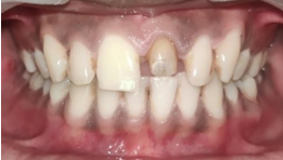
A young male patient aged 19 years, reported to the department of prosthodontics, with the chief complaint of broken upper front teeth due to trauma.
Brief History revealed that patient had undergone root canal treatment 3-4 years back with 21.
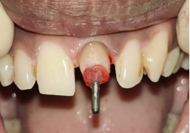
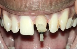
On clinical examination, 3-4 mm tooth structure present supra-crestally.
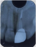
Radiographic examination revealed a wide & flared root canal.
After clinical and radiographic evaluation, a decision was made to restoring the lost tooth structure with the anatomic post and core followed by zirconia crown with 21.
Procedure
The preparation of the post space was carried out following all standard principles, utilizing PEESO reamers (MANI, Japan).
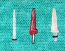
A size 4 glass fiber post (Mailyard fiber post) was chosen and assessed for fit, revealing a poor fit within the canal. Consequently, the decision was made to construct an anatomical post using an indirect extraoral technique.
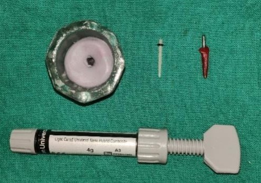
To execute this, an old, worn-out diamond bur (MANI, Japan), pattern resin (GC), condensation silicone (Zermack, Zetaplus), glass fiber post (Mailyard fiber post), and composite resin (Prevest Fusion Universal Composite) were employed.
The canals were dried and coated with petroleum jelly. Subsequently, pattern resin was conventionally mixed and applied to the old, worn-out bur.
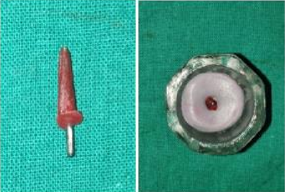
Pattern resin (GC) was conformed to the bur, and this bur was promptly inserted into the root canal, with shaping of the coronal portion before setting.
After setting, the pattern resin post space impression was extracted from the prepared post space.
Condensation silicone putty (ZERMACK ZETAPLUS) was mixed and positioned inside a glass dappen-dish, and the pattern resin post space impression was placed in this putty.
Once the putty had set, the bur was removed to obtain the silicone replica of the prepared post space.
Silane (Ultradent, USA) was applied to the intended fiber posts for one minute.
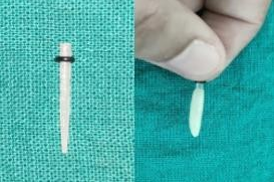
A nano-hybrid composite resin (Prevest Fusion Universal Composite) was shaped onto the post, which was then loosely inserted into the silicone replica of the prepared post space.
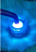
Initial light curing for 20 seconds from above was followed by additional curing for 20 seconds after retrieval from the silicone replica.
The prepared anatomical post was checked intraorally for proper fit and retention, demonstrating a snug fit and secure retention in the post space.
The prepared anatomical post was cemented using dual-cure resin cement (Prevest Fusion Ultra D/C).
Core buildup was performed over the cemented post, and tooth preparation was carried out to accommodate an All-Ceramic zirconia crown.
Gingival retraction was achieved using Sure-endo 000 retraction cord, and a silicone impression (Dentsply Aquasil soft impression material) was created.
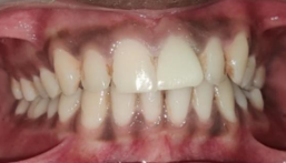
A CAD-CAM Zirconia crown was fabricated and cemented using dual-cure resin cement (Prevest Fusion Ultra D/C).
Periodic follow-ups were conducted.
Discussion
Due to trauma during early tooth development, the tooth in question exhibited a wide and flared root canal with thin radicular dentin. The current scenario, characterized by a wide and flared canal, deemed it suitable for the creation of an indirect anatomical post. The introduction of a conventional fiber post into the canal would have necessitated a substantial layer of luting cement to address the spaces between the loosely fitting post and the canal walls. This approach posed a risk of adhesive failure and post debonding.7 Opting for a well-fitted post tailored to the canal shape allowed for the use of a thin, uniform layer of cement, thereby enhancing retention.
Some authors have proposed the use of accessory posts to fill the space around the main glass fiber post. However, this approach does not effectively reduce cement space, as multiple posts introduce numerous voids. In the direct anatomical post technique, the fiber post is relined in the root canal, replacing the resin cement with composite resin, known for its superior mechanical and physical properties. While relatively straightforward, this technique has limitations, such as being time-consuming and tedious.
Certain authors 8, 9, 10, 11 have advocated for restoring flared root canals with composite resin to reduce canal width. However, challenges in achieving adequate curing at the deepest regions of the canal wall may affect the material's properties 12 and its bonding to the adhesive layer. In contrast, the indirect anatomical post addresses this issue, as the composite resin attached to the fiber post is initially cured inside the silicone replica and further cured before the luting procedures.
Although indirect anatomical posts boast superior mechanical properties, their fabrication is often considered time-consuming and expensive due to the required laboratory step. This clinical report introduces an innovative dental technique to overcome these drawbacks and facilitate the fabrication of indirect anatomical posts in a straightforward manner.
The advantages of this technique include
Its independence from additional laboratory steps.
Cost-effectiveness.
Chair-side applicability.
Reduced time consumption.
Minimal material requirements.
Superior mechanical properties compared to direct anatomical posts.
In light of the growing preference for esthetic metal-free restorations, the use of indirect anatomical posts emerges as a practical solution for managing wide and flared root canals in routine clinical practice.
Conclusion
Applying an innovative, practical, cost-effective, and expeditious technique involves utilizing indirect anatomical posts in wide and flared root canals. This method is applicable for both direct and indirect esthetic restorations, with the goal of enhancing the bond strength between the fiber post and the root canals, while concurrently reducing the common risk of fractures associated with cast-metal posts.

