Introduction
A kind of optical microscope termed a stereo, stereoscopic, or dissecting microscope is employed to observe a sample with low magnification. It operates by bouncing light on an object's surface rather than passing it through. To provide slightly different viewing angles to the left and right eyes, the device uses two independent optical pathways with two objectives and eyepieces. By employing this arrangement, the sample under examination can be seen in three degrees.
When recording and learning solid samples with complex surface topography—where a three-dimensional viewpoint is needed for detail analysis—stereomicroscope surpasses macro photography.
When analyzing the surfaces of solid substances or conducting up-close activities like dissection, microsurgery, watchmaking, circuit board manufacture or inspection, or analyzing fracture surfaces as in fractography and forensic engineering, the stereo microscope is often used.
Discussion
A compound microscope with two eyepieces and a Bino viewer should not be confused with a stereo microscope. With such a microscope, the two eyepieces serve to improve viewing comfort as both eyes see the same image. However, the image produced by such a microscope resembles the image generated by a single monocular eyepiece. 1
History
The first optically practical stereomicroscope was created in 1892 and produced by Zeiss AG in Jena, Germany, which came on the market in 1896.
Son of famous sculptor Horatio Greenough Sr., American scientist Horatio Saltonstall Greenough grew up in Boston's elite. He went to France to pursue a career in science instead of feeling the burden of having a job for a living. He was influenced by the new scientific concepts of the day, notably experimentation, at the marine observatory in Concarneau on the Bretton coast, which was run by Georges Pouchet, the previous head of the Museum National d'histoire Naturelle.
During Greenough's time at Con Carneau, interest in performing research on live and developing organisms was revived, though zoologists, anatomists, and morphologists had previously focused primarily on the dissection of prepared and dead specimens. Scientists may then observe the process of embryonic development rather than seeing it as a collection of two-dimensional, petrified specimens. A new microscope was needed to provide images that accurately depicted the relative size and three-dimensionality of developing invertebrate ocean embryos. Encouraged by his colleague Laurent Chabry's work at Concarneau to create complex mechanisms to turn and regulate the living embryo, Greenough moved ahead and built his instrument.
Building on Charles Wheatstone's recent discovery that binocularity is the source of depth perception, Greenough built his tool with the idea of stereotyping in mind. 2
Differentiating from normal microscope
Compared to a compound light microscope, a stereo microscope often uses reflected illumination as compared to transmitted (diascopy) illumination, and light that is reflected from an object's surface in contrast to light that is transmitted through it. Specimens that would be too dense or too opaque for compound microscopy can be studied via the object's reflected light. Although transmitted illumination is not focused through a condenser in most systems, unlike compound microscopes, some stereo microscopes can also illuminate a thing with transmitted light. This usually happens by placing a bulb or mirror behind a transparent stage beneath the object. 3, 4
Darkfield microscopy can be done with either transmission or reflected light using stereoscopes which have been properly fitted with illuminators. The key features of this sort of microscope are a great working distance and depth of field. Resolution has an inverse relationship with these features: the greater the resolution (i.e., the separation between two nearby spots at which they can be determined to be distinct), the smaller the working distance and depth of field. Although the magnification is typically much lower, some stereo microscopes offer a practical magnification of up to 100×, which is comparable to a 10× objective and 10× eyepiece in a typical compound microscope. This is roughly a tenth of what a typical compound optical microscope can detect.
When employing fiber-optic illumination, as will be discussed later, the large working distance at low magnification can help inspect large solid objects such as fracture surfaces. It is very simple to alter these data to find the points of interest. 5
Magnification
Stereo microscopes have a pair of primary types of magnification processes. A particular type is fixed magnification, in which a matched pair of objective lenses with a preset degree of magnification provides the primary magnification. The other is zoom or pancreatic magnification, which may differ the magnification constantly across an established range. With the use of auxiliary objectives, zoom appliances can boost magnification further by a predetermined factor. In addition, by changing out the eyepieces in fixed and zoom systems, the overall magnification can be adjusted. The "Galilean optical system" is a system attributed to Galileo that collapses between fixed and zoom magnification systems. It makes use of a set of fixed-focus convex lenses to provide a fixed magnification, but it has an important difference in that the same optical components in the same spacing will give a different, though still fixed, magnification if physically inverted. It allows two sets of lenses to provide four magnifications on one turret, three sets of lenses to provide six magnifications, and one set of lenses to provide two different magnifications. Practical usage shows that these Galilean optics systems are just as helpful as a far more expensive zoom system, with the added benefit of making one aware of all of the magnification utilized. 6, 7
Illumination
Fiber-optic sources of light tend to be employed to provide the intense illumination that small specimens require, especially at high magnifications. Halogen lamps, which generate a lot of light for a specific amount of power input, are used in fiber optics. Though they frequently require cooling to reduce high temperatures from the bulb, the lamps are small enough to be placed close to the microscope with comfort. The operator has an abundance of flexibility in choosing ideal lighting conditions for the sample thanks to the fiber-optic stalk. The sheath surrounding the stalk is elastic and may be slipped into any desired position. When the highlighted end of the stalk gets near the specimen, it is typically not visible and doesn't interfere with the image under the microscope. Fiber-optic lights are perfect for oblique lighting, which is frequently needed for fracture surface examination to bring out the surface's features during fractography. For the same specimen, several of these kinds of light stalks can work, increasing the illumination. High-power LEDs, which are far more energy efficient than halogen lights and can produce a spectrum of colors of light, are a more recent development in the lighting for dissecting microscopes. This makes them useful for fluorophore analysis of biological samples, which is impossible with a halogen or mercury-based light source. 8
Digital microscope
Digital stereo microscope overview
A stereo microscope, also known as a dissecting microscope, is designed for low-magnification observation of samples.
Unlike compound microscopes, which use transmitted light, stereomicroscopes rely on reflected light from the surface of the specimen.
They provide a three-dimensional visualization by presenting slightly different views to the left and right eyes.
Stereomicroscopy is essential for studying solid specimens with complex surface topography, such as dissection, microsurgery, watch-making, circuit board manufacture, inspection, and fracture surfaces (as in fractography and forensic engineering).
These microscopes are widely used in manufacturing industries for manufacture, inspection, and quality control.
The stereo microscope should not be confused with a compound microscope equipped with double eyepieces and a bino viewer, where both eyes see the same image, providing greater viewing comfort but no different image from that obtained with a single monocular eyepiece.9, 10
Integration of digital displays
Some modern stereomicroscopes come equipped with integrated digital cameras.
These cameras capture magnified images of the specimen.
The images can be displayed on a high-resolution monitor or other digital screens.
This integration allows researchers, educators, and professionals to view and analyze specimens without the need for traditional eyepieces.
It’s particularly useful for collaborative work, teaching, and documentation.
Benefits of digital displays
Real-time Observation: Researchers can observe specimens in real-time on a large screen, making it easier to share findings.
Image Capture: Magnified images can be captured and saved for further analysis, reports, or educational purposes.
Three-Dimensional Imaging: Digital displays enhance the three-dimensional effect, aiding in depth perception.
Measurement and Annotation: Some systems allow measurements and annotations directly on the displayed image.
STereo microscope parts and functions
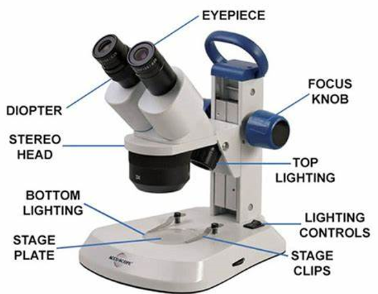
The three main components of it are the body, the focus block, and the viewing head/body.
The body/viewing head of the microscope is where the optical components are located in the upper section.
Optical parts
The eyepiece lenses are located at the microscope's top. The standard eyepiece has a 10X magnification.
The optional eyepiece comes with a power range of 5X to 30X.
The eyepiece tube, which is located directly above the objective lens, holds the eyepieces in place.
Stereo microscope types
Stereo fixed microscope
Since it uses two objective lenses to provide fixed magnification, it is also known as a fixed stereo microscope. The lens's capability defines the set degree of magnification. You have to switch out the eyepiece to increase the magnification.
Stereo turret microscope
One of the mounting possibilities that it comes with is the turret style. This kind of setup shows an extra objective lens that you can turn depending on your viewing angle.
The turret mounting may be readily adjusted by the viewer to adjust the magnification. Because it is less costly than other kinds of stereo microscopes, it is preferred by many.
Differences between – Stereo vs Compound microscope
Optical route
Nature
Expansion
Low amplification is used with a stereo microscope.
Compound microscopes may amplify images up to 1000 times.
Light
Stereo microscope: Reflected light is used to view the specimen.
Compound microscope: The object lets light pass through it.
Uses
Stereo microscope is used to look at a solid material's surface.
Compound microscope is used to look at tiny objects 14
Dissecting microscopes, also known as stereomicroscopes, are instruments that connect the powerful world of compound microscopes with the human eye. Their ability to provide a distinct viewpoint for examining and working with small specimens makes them indispensable instruments in many scientific domains.
Three-dimensional (3D) visualization
Stereomicroscopes utilize independent optical channels for each eye to provide a stereoscopic perception, compared to compound microscopes, which establish a flat viewpoint. In order to perform operations like dissection and microsurgery, viewers must be able to recognize depth and spatial relationships within the specimen.
High resolution at low magnification
Stereomicroscopes are most appropriate for observing larger, transparent objects at magnifications between 10 to 50x, a level lower than compound microscopes. This allows for an in-depth examination of surface features and beyond structures, especially when combined with their high resolution.
Large working distance
The distance from the objective lens and the subject in a stereomicroscope is larger than that of a compound microscope. It gives space to utilize devices such as forceps and microscalpels, especially helpful in dissections and other difficult procedures.15
Note
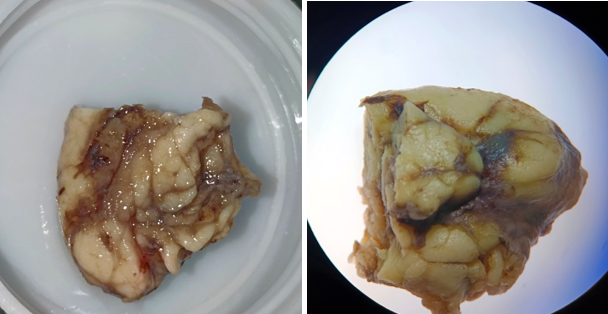
Under stereomicroscope lymph node are viewed in low power magnification. The maximum view of the stereo microscope is 100 X. With the stereomicroscope the lymph node structures can be easily identified and also this can be used to identify and also this can be used to identify the solid structures, gross specimens, microleakage and depth of filling material in endodontics
Conclusion
Stereomicroscopes provide a special and useful viewpoint for applications which require for the handling and observation of small things. Because of their ease of use and capacity to provide a magnified 3D picture, they are essential instruments in an array of scientific domains and educational environment.


