- Visibility 17 Views
- Downloads 4 Downloads
- DOI 10.18231/j.idjsr.2023.018
-
CrossMark

Laser-assisted excision of peripheral ossifying fibroma- A case report
Introduction
Localized gingival growths are one of the maximum often encountered lesions inside the oral cavity which simulate some scientific & histopathologic features. Peripheral Ossifying Fibroma (POF) is one of the commonest lesions of the oral cavity. It accounts for 3.1% of all oral tumors and for 9.6% of gingival lesions.[1], [2] POF is a focal, reactive, non-neoplastic connective tissue lesion that often arises from the interdental papilla and develops on gingiva as a response to irritants such as trauma, microorganisms, plaque, calculus, and restorations. [3], [4] It has a tendency to arise especially at 20-30 years of life, with the highest prevalence between 10 and 19 years. Those lesions might also rise up due to such irritants as trauma. About 60% of those tumors occur within the maxilla and 50% of all instances of maxillary POF are located in the incisors and canine areas.[5] Incidences of recurrence of POFs are observed to be 8.9–20% because of incomplete removal of the lesion, incomplete removal of local irritants, and inaccessible approach for the surgical excision due to the complex location of POF usually present at interdental regions. In order to avoid this, deep excisions are favored. [6] This case file depicts a case of POF in a 31-year-old lady in the maxillary anterior area. The lesion was surgically excised followed by a follow-up and till 6 months there was no recurrence.

Case Report
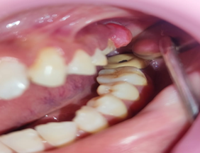
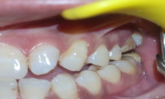
A 31-year-old woman visited the OPD of KD Dental College and Hospital, Mathura with the chief problem of a painless overgrowth in her left maxillary posterior vicinity for the past 6 months. Intraoral examination showed a solitary, sessile gingival overgrowth extending mesiodistally from the mesial surface of #26 to the distal surface of #27. Cervico-inicisally, the growth prolonged from the attached gingiva till occlusal surfaces of the maxillary left 1st and second molars. The coloration of the growth looks similar to that of the attached gingiva with an extra reddish-crimson presentation on the cervical element. [[Figure 1]] On palpation the swelling turned into non-tender, firm, non-compressible, and related to moderate bleeding on provocation. The growth changed into an oval of approximately 12 mm × 4 mm in length. Records revealed that the swelling initially began with the dimensions of a pea and grew hastily to the prevailing size. The affected person complained of difficulty in speech and mastication due to the swelling. The affected person noticed similar growth in the maxillary posterior location a year back which was excised via a scalpel and noticed a comparable recurrence in the ultimate 6 months. A radiographic exam revealed erosive bone changes around the interdental bone of #26 & #27. The differential diagnosis suggested was Pyogenic granuloma, fibroma, and peripheral ossifying fibroma & provisional diagnosis of inflammatory enlargement was made.
Surgical Procedure
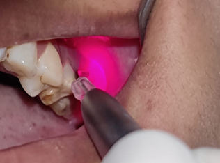
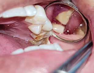
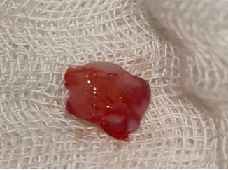
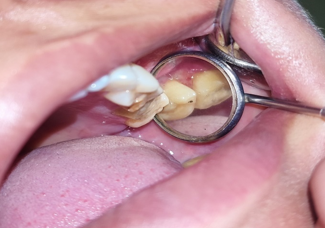
The affected person’s consent was taken prior surgery. Anesthesia was administered, and with the help of LASER, the lesion was excised followed by the curettage and reshaping of gingival tissues. [[Figure 2]] Periodontal dressing was given over the surgical site. [[Figure 5]] Analgesics & antibiotics had been prescribed to the affected person and postoperative instructions have been given. Mouth rinse using 2% Povidone-Iodine mouth rinse was instructed for 10 days and was recalled after 10 days for re-evaluation and elimination of the dressing. The excised tissue was positioned in 10% impartial buffered formalin and sent for histopathological evaluation. Microscopically the tissue consisted of a mild to moderate to distribution of persistent inflammatory cells inside the fibrovascular connective tissue stroma and hyperplastic para-keratinized stratified squamous epithelium. A couple of abnormal bony trabeculae showing the presence of osteocytes in lacunae will be visualized. After correlating the clinical, radiographic, and histopathologic functions, POF was considered as the final diagnosis. At 10 days post-operative visit, periodontal dressing was removed, and healing was satisfactory. Regular follow-ups were done and no signs and symptoms of recurrence were noticed till 6 months.
Discussion
Menzel in 1872 first defined the Ossifying fibromas and Bernard Law Montgomery in 1927 gave its terminology. Peripheral ossifying fibromas are in addition labeled as significant and peripheral. The central portion arises from the endosteum or the periodontal ligament (PDL) adjacent to the root apex while the peripheral type takes place at the soft tissues overlying the alveolar mucosa. POF is a solitary, gradual-growing nodular mass this pedunculated or sessile. It generally occurs within the gingival papilla between neighboring teeth. But the etiopathogenesis of POF is foreseeable, it is believed to be originating from cells of pdl due to the subsequent reasons –
Frequently inside the gingival interdental papilla,
Its closeness to the gingival to pdf,
The fibro-cellular response in pdl and
The presence of oxytalan fibers within the mineralized matrix of some lesion. [7], [8], [9]
Predisposing elements of POF consist of trauma to the gingiva, plaque accumulation, ill fitting dental appliances and crowns, masticatory forces, defective restorations, and many others. Differential diagnoses of POF are traumatic fibroma, peripheral odontogenic fibroma peripheral giant cell granuloma, and pyogenic granuloma. [10] Peripheral giant cell granuloma suggests a bluish or purple discoloration that is not found in POF. Peripheral odontogenic fibroma is an extraordinary neoplasm that arises from odontogenic epithelial rests in the periodontal ligament or attached gingiva itself. Histologically, peripheral giant cell granuloma includes giant cells and peripheral odontogenic fibroma includes odontogenic epithelium and dysplastic dentine. Those features are absent in POF. Pyogenic granuloma gives as a tender, friable nodule, small in length that bleeds and has an inclination to hemorrhage. [11], [12], [13]
Management requires proper surgical intervention that guarantees deep excision of the lesion together with the periosteum and affected periodontal ligament. Thorough root scaling of adjoining teeth and/or elimination of different sources of irritants should be executed. Treatment modalities for POF include the elimination of local irritants and surgical excision by the usage of a scalpel, lasers, or electrosurgery. Recurrence chances of POF vary from 8.9 % to 20 %. The most common cause of recurrence is the partial or incomplete removal of the lesion. [14], [15], [16]
Conclusion
POF is a benign, solitary slowly progressive lesion that is frequently misdiagnosed as pyogenic granuloma. No matter the surgical method employed, it's very crucial to remove all the etiological factors and the tissue has to be histologically tested for confirmation. Careful clinical and radiographical examination correlated with histopathological findings is required for the final diagnosis. Consequently, histopathological examination is required to verify the diagnosis. POFs are treated through entire surgical excision and gingival curettage, accompanied by precise oral hygiene maintenance. Patients need to be followed up for an extended time period because of high relapse chances.
Source of Funding
None.
Conflict of Interest
None.
References
- JN Kenney, GE Kaugars, LM Abbey. Comparison between the peripheral ossifying fibroma and peripheral odontogenic fibroma. J Oral Maxillofac Surg 1989. [Google Scholar] [Crossref]
- JD Walters, JK Will, RD Hatfield, DA Cacchillo, DA Raabe. Excision and repair of the peripheral ossifying fibroma: A report of 3 cases. J Periodontol 2001. [Google Scholar] [Crossref]
- T Farquhar, J Maclellan, H Dyment, R D Anderson. Peripheral ossifying fibroma: A case report. J Can Dent Assoc 2008. [Google Scholar]
- SK Kumar, S Ram, MG Jorgensen, CF Shuler, PP Sedghizadeh. Multicentric peripheral ossifying fibroma. J Oral Sci 2006. [Google Scholar] [Crossref]
- DG Gardner. The peripheral odontogenic fibroma: an attempt at clarification. Oral Surg Oral Med Oral Pathol 1982. [Google Scholar] [Crossref]
- CS Miller, RG Henry, DD Damm. Proliferative mass found in the gingiva. J Americ Dent Assoc 1990. [Google Scholar]
- VJ Barot, S Chandran, SL Vishnoi. Peripheral ossifying fibroma: A case report. J Indian Soc Periodontol 2013. [Google Scholar]
- SK Kumar, S Ram, MG Jorgensen, CF Shuler, PP Sedghizadeh. Multicentric peripheral ossifying fibroma. J Oral Sci 2006. [Google Scholar] [Crossref]
- S Chhina, A Rathore, A Puneet. Peripheral ossifying fibroma of gingiva: a case report. Int J Case Rep Images 2011. [Google Scholar]
- M Agarwal, G Bhatia, V Wadhawan, KV Kumar. Mucoepidermoid Carcinoma- A chance finding in gingival overgrowth. Clin Adv Perio 2018. [Google Scholar]
- LR Eversole, AS Leider, K Nelson. Ossifying fibroma: a clinicopathologic study of sixty-four cases. Oral Surg Oral Med Oral Pathol 1985. [Google Scholar] [Crossref]
- N Rana, S Joshi, M Kaushik, P Singh. Peripheral Giant Cell Granuloma. Int J Creative Res Thoughts 2022. [Google Scholar]
- G Jain, R Arora, A Sharma, R Singh, M Agarwal. Irritation fibroma: Report of a case. J Cur Res in Sci Med 2017. [Google Scholar] [Crossref]
- A Sharma, S Singh, M Kaushik, S Khattri. Puberty induced Gingival enlargement- A clinical case report. J Adv Med Dent Sci 2021. [Google Scholar]
- M Tamarit-Borrás, E Delgado-Molina, L Berini-Aytés, C Gay-Escoda. Removal of hyperplastic lesions of the oral cavity. A retrospective study of 128 cases. Med Oral Patol Oral Cir Bucal 2005. [Google Scholar]
- PS Koodakandi, A Shigli, DD Shirol. Peripheral ossifying fibroma: A clinical report. Contemp Clin Dent 2010. [Google Scholar]