Introduction
Since ancient times, maxillary arch expansion has been investigated. Due to the promising outcomes of numerous research, doctors now use both slow and quick palatal expansion tools to widen the transverse dimension of the constricted maxillary arch. Emerson C. Angell is credited as being the father of fast maxillary expansion since he described his first instance of successfully splitting the maxilla with a jack screw appliance in 1860. Farrar and Clark Godard (1893), emphasised the value of transverse palate expansion together with opening up of the mid palatal suture. Latha m held the view that growth at the midpalatal suture stops at the age of three years, however Bjork and Skieller's work using implants in the year 1974 disproved Latham's theory and proved that growth at the midpalatal suture can last up to 13 years. Dentoalveolar expansion, which involves the use of appliances to widen the palate in a transverse orientation, is another name for slow maxillary expansion. Despite the enlargement being entirely dental, some skeletal alterations are visible. The patient's age and the amount of force used are the only factors that affect the effects of skeletal alterations vs dental changes. It wasn't until 1978, however, when Hicks with his cephalometric study demonstrated the efficacy of slow palatal extension by using a fitting split acrylic plate to widen the maxillary arch. 1
Classification of Slow Maxillary Expansion Appliances1
Slow maxillary appliances can be broadly classified as follows:
Advantages of SME 1
It continuously distributes physiologic force, until the necessary expansion is attained
There is minimum tipping of anterior teeth.
Least strain is exerted on anchored teeth.
The appliance is light and comfortable to the patient.
Relapse tendencies are less.
It can be used for sufficient retention after the expansion.
Time required for retention is less.
Throughout its use, it takes little adjustment and makes adjustments simple when needed.
Maintenance of sutural integrity and the reduced stress loads within the tissues
Do to lighter force application, patient experiences less discomfort and pain.
Age Factor on Treatment Outcome 1
According to Proffit, 1-2 lbs of forces are sufficient to cause expansion in younger children. However, in comparison to skeletal alterations, dental changes are more frequently seen in teenagers. According to Hicks, greater activation offered limited expansion whereas forceful activation caused the anchored teeth, primarily the molars, to tip, producing the most dental alterations in adults. In his investigation, he applied a 2 lb force with predicted expansion rates of 0.5 to 1.0 mm each week, yet he managed to widen the maxillary arch by 3.8 to 8.7 mm while receiving therapy. He claimed that in children aged 10 to 11 years, skeletal alterations accounted for 24 to 30 percent of the overall rise in arch width, whereas in patients aged 14 to 15 years, the expansion rate was only 16 percent.
Effects of Sme On Mid-Palatal Sutures1
The opening of the mid-palatal suture occurs when sutural integrity is maintained during maxillary remodelling, according to Storey et al in 1973. In 1977, Ekstrom et al. demonstrated that SME provides stronger sutural stability, greater reparatory response, and less traumatic disruption than fast expansion of sutures. Bell et al in 1982 claimed that compared to the disruptive character of rapidly expanded maxillary segments, the pace of midpalatal suture separation by moderate expansion systems evidently provides a more physiologically bearable response by the sutural elements. The amount of orthodontic movements is increased by modest expansion techniques, according to Moyers et al in 1974, since the tensile strength of the suture parts is not exceeded. In a comparison study of slow and rapid palatal expansion performed in 1982 by Zachrisson et al., it was found that while periodontal breakdown on the buccal surfaces of the back teeth occurred in both groups, the incidence of attachment loss was higher in the rapid palatal expansion group.
Appliances Used To Produce Slow Maxillary Expansion
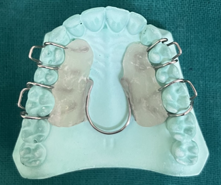
Coffin spring
In the year 1875, Sir Walter Coffin invented the coffin spring. The appliance comprises of an omega-shaped wire put in the mid-palatal area and an adam's clasp in the first premolars and first molars on both sides. The appliance's components are embedded within an acrylic base plate and constructed from 1.2 mm stainless steel wire. The appliance is largely suggested to cause dentoalveolar alterations in cases of unilateral or bilateral crossbite, cases requiring lateral expansion, cases requiring antero-posterior expansion, and cases where the space required is less than 3 mm. However, some amount of skeletal changes can also be brought about in mixed dentition period if proper retention protocol is maintained.2
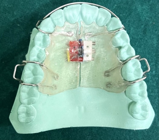
Active plate1
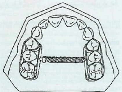
Pierre Robin first proposed the idea of an active plate in 1902. He fabricated a split acrylic plate with a screw in the middle to allow the arch to expand. The acrylic base of the active plate acts as a base for screws or springs to be incorporated and clasps to be hooked to. The working parts of this detachable appliance are the expansion screws. According to Proffit, the majority of screws open 1mm every full rotation, translating to 0.25mm of tooth movement per quarter turn.The active plate are most useful when few millimetres of space is required (1.5-2mm per side). (Figure 2)
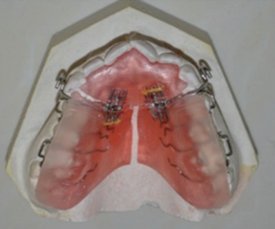
Plate1
It is an active type of removable expansion device similar to the bite plate, with Adams clasps acting as an anchorage or retentive component in the premolar and molar region. The retentive arm is inserted in the acrylic, and the labial bows are placed in the anterior region. Two jack screws are positioned between the front and posterior halves of the acrylic plate, splitting it into a Y shape and applying a distalizing force. When the jackscrews are activated, the buccal segment teeth experience a distalizing force, and the anterior palatal contour and maxillary incisors experience a reciprocal force. The jackscrews are activated alternately to prevent the incisors from tipping labially and from dislodging the entire device out of place. The Y plate is indicated in patients with first premolars erupted, giving increased anchorage, upright incisors and where no extensively bodily movement are required. (Figure 3)
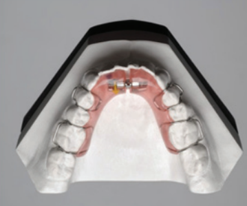
Shwartz appliance1
In 1966, Shwartz developed the Shwartz appliance. It is mostly constructed for the mandible and is a removable expansion plate. The appliance is indicated during the phase of mixed dentition. The acrylic plate that serves as the appliance's main structural element has a midline split, one or two expansion screws, and does not cap the occlusal surface or the incisal borders. The device is secured with ball or Adam's end clasps and has a labial bow as well. The Schwarz appliance may be helpful for patients with short arch lengths and/or posterior teeth with abnormal lingual inclinations. The activation of the midline screw, which causes the Schwarz appliance to gradually expand, only causes the posterior teeth to point laterally .This is followed by rapid maxillary expansion which would stabilize mandibular dentoalveolar position during the retention period. (Figure 4)
Quad helix
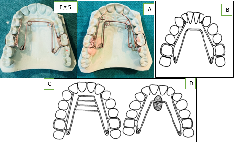
Robert ricketts first introduced it in 1975. It is an upgrade of the W arch appliance. The indications include the following: a) All crossbites require upper arch extension; b) Cases of crowding necessitating mild expansion. c) Class II cases requiring molar distal rotation; d) Class III instances requiring maxillary arch surgery; e) situations involving tongue thrusting; and f) patients with cleft lip and palate. The .038 Elgiloy, No. 4 Gold, or 1 mm S.S. that makes up the quad helix is used. The distal surfaces of the canines are level with the anterior bridge. The palate is where the anterior helices are located. Distal to the first molars is where the posterior helices are located. These slope in a straight line with the palatal surface. Prior to cementation, the activation is completed. To separate the midpalatal, Ricketts advises 500 mg force.1 (Figure 5)
Modifications
The quadhelix appliance may be modified in a number of ways. Modifications usually fall into one or more of the following categories:3
Increased number of helices: Extra helices may be introduced to procline the upper incisor teeth in order to allow for antero-posterior extension. If space maintenance and molar derotation rather than upper arch extension are the goals of treatment, a palatal button may be exchanged for a palatal button with fewer helices and anterior helices omitted.3 (Figure 5 A)
By convention, a device with only posterior helices that is positioned in the lower arch is referred to as a "bihelix.".3 (Figure 5 B)
Addition of habit breaking auxiliaries, Bench et al. (1978) describe the addition of tongue spurs and grids to discourage digit or tongue sucking habits.3 (Figure 5 C) Figure 5 A
Warch
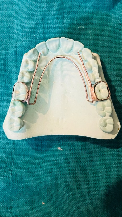
Robert ricketts first created the W arch appliance in 1975. It is a fixed device in the form of a horseshoe that is attached to the molar bands on either side. In the maxilla and mandible, where modest growth is favoured, it is advised. The construction of the lingual arch is done 1-1.5 mm away from the palate in order to prevent any harm to the soft tissues. The W arch appliance is activated by opening the apices of the appliance. The arch is adjusted based on whether anterior or posterior expansion is required. The appliance is preactivated before insertion by opening up the apices by 3mm in order to give the proper force levels.1 (Figure 6)
NiTi Expander
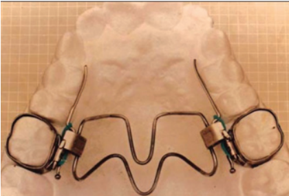
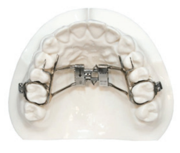
Wendell introduced the Nickel Titanium Palatal Expanders (Figure 7). It produces constant expansion forces that are ideal. The main component is constructed of a NiTi alloy that has been thermally activated, while the rest of the parts are made of stainless steel. The expander merely needs an extra lingual sheath on the molar bands in order to be utilised in conjunction with traditional fixed appliances. The appliance's functionality results from the shape memory and transition temperature properties of nickel titanium. enlargement of the maxilla slowly using nickel titanium. The transition temperature for the nickel titanium component is 94° F. The expander is too stiff to flex for insertion at room temperature. The primary component of the expander softens after being chilled, making manipulation easier. stiffens and starts to take on its original shape after being put. Only 350 gms of force is applied every 3 mm of expansion, and the nickel titanium alloy maintains relatively constant force levels when the expander deactivates.4
Spring Jet
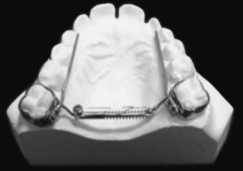
The molar bands are soldered to or otherwise connected to the spring jet's active components (Figure 8). To ensure that forces pass close to the centre of resistance of maxillary teeth, the telescopic unit is positioned up to 5 mm from the centre of the molar tubes. However, it should be 1.5 mm away from the palatal tissue. In the mixed dentition, 240 gm of force is used, and 400 gm in the permanent dentition. The lock screw must be moved horizontally along the telescopic tube to activate. A ball stop on the transpalatal wire allows the spring to be compressed.5
Minne Expander
Minne Expander is a fixed, slow maxillary expansion appliance cemented to the first permanent molars and first premolars. The palatal compressed-coil spring is activated in order to improve maxillary width. According to Hicks 1978, the Minne-expander appliance spring applies forces of up to 10 N which is upto 2 pounds. the expansion of the Minne-expander is 0.125 mm for each incremental activation. The maxillary sutures are less affected by it, and as a result, they recover and are repaired during the expansion treatment, giving it a more physiologic appearance. Poor oral hygiene maintenance is a drawback of this appliance.1 (Figure 9)
Spring Loaded Expander
Leone debuted the spring-loaded expander (SLE) in 2003. Using modest continuous pushes, the SLE, a novel expansion device, causes gradual palatal expansion. Patients with finished growing should use the appliance. The control over the spring allows them to achieve precise force levels. SLE has the ability to generate either a 500g or an 800g of force, depending on the requirement for expansion. The appliance consists of bands surrounding the molar with screw attached to the centre. The spring provides a continuous force, sufficient to promote a dentoalveolar remodelling that is biologically ideal and biomechanically controlled. In order to prevent disassembly in the event of excessive activation, the screw incorporates a self-stop mechanism near the end of expansion. Every six weeks, the gadget receives an average of 4 to 8 activations (0, 4-0, 8 mm). The force applied to the dental structures will remain consistent in intensity regardless of the number of activations (500 or 800g.). Since the screw will become inactive once it has expanded by the predetermined amount, there is no chance of over-expansion. However, fast maxillary growth can also be accomplished utilising SLE by altering the activation sequence.1 (Figure 10)
Discussion
The element of treatment that has been most closely studied in relation to changes in maxillary expansion is the pace of expansion. Slowly expanding procedures, such those that employ lingual arch wire equipment with expanding capabilities (Fig. quad helix), use force systems ranging from a few ounces to around 2 pounds.6 As the tensile strength of the suture elements is not exceeded, the modest expansion operations enhance the percentage of orthodontic motions. However, sluggish maxillary expansion in both humans and primates has been linked to orthopaedic separation of the maxillary segments.7 Skieller estimated that the orthopaedic separation of the midpalatal suture was responsible for around 20% of the rise in arch width when serial frontal cephalograms taken during gradual expansion treatment (0.5 mm per week) were analysed using established reference standards.8 Hicks obtained maxillary arch width gains of between 3.8 and 8.7 mm throughout treatment utilising 2-pound pressures with expansion rates of 0.4 to 1 mm per week. Hicks calculated that the skeletal changes accounted for between 24 and 30 percent of the total increase in arch width in patients aged 10 to 1l and 16 percent in those aged 14 to 15 using nonanatomic reference points.7 Histologic findings, reported in conjunction with slow expansion procedures, suggest that sutural separation occurs at a rate which allows the maintenance of tissue integrity during adjustments to the maxillary repositioning and remodeling. 9 According to Ekstrom and colleagues, the slowly expanding suture typically takes approximately 30 days to become well organised by mineralized tissue and takes three months to become established.10 Storey argues that moderate suture growth of 0.5 to 1.0 mm per week, as opposed to rapid expansion, enables "physiologic sutural alterations" with less traumatic disruption, a stronger reparative reaction, and more sutural stability.9 Reduced evidence of abutment tooth tipping and the accumulation of residual stress loads inside the larger segments following slow maxillary growth are two further comparable impacts that have been observed. After gradual expansion treatments, doctors have empirically noticed sutural integrity preservation, a decrease in stress loads within the tissues, and a lower chance of skeletal recurrence.11 Retention periods of 3 months or less appear adequate in allowing sutural regeneration and stabilization of slowly separated maxillary segments.7, 10
Early intervention appears to facilitate substantial increases in maxillary arch width in addition to increased response to maxillary expansion through the use of less complicated and lower-force expansion devices. The relative quantitative increments obtained with low-force, moderate expansion treatments are less skeletally important but still favourably contrast with the qualitative orthopedic/orthodontic changes observed following rapid expansion operations in prepubertal age groups. The pace of midpalatal suture separation by slow expansion systems appears to provide a more physiologically tolerable reaction by the sutural elements, in contrast to the disruptive character of rapidly expanded maxillary segments. Improved tissue integrity maintenance in slowly increasing sutural elements has been associated with enhanced stability and a decreased risk of recurrence throughout the remodelling of the maxillary complex. The slow expansion protocol's ensuing retention durations of 1 to 3 months are much shorter than the 3- to 6-month regimens advised to maintain rapidly developed maxillary arches.12 While the research seems to support early correction utilising a gradual expansion method, specific factors must be taken into account when choosing an expansion protocol that will best influence the scope and quality of the expansive alterations. Even while older individuals may need the greater force systems of quick expansion procedures or surgical intervention to accomplish palatal separation, fixed lingual arch wire expansion appliances may still be an efficient way to widen the maxillary arch during the deciduous and mixed dentitions.13, 14
Conclusion
The best technique to gain space in the past was believed to be tooth extraction. However, there has long been debate about whether the dentition and related structures can survive after teeth are removed. One of the easiest and safest methods to expand your space is through an arch. Researchers are leaning toward both skeletal and dental consequences of slow maxillary palatal expansion due to the aggressive nature of rapid palatal expansion on tissues, even though both quick and slow maxillary arch growth have demonstrated to produce long term stability. However further clinical trials have to be carried out to discuss the effects of SME on dentition.

