- Visibility 87 Views
- Downloads 5 Downloads
- DOI 10.18231/j.idjsr.2020.026
-
CrossMark
- Citation
Fabrication of ocular prosthesis step by step procedure : Case report
- Author Details:
-
Jyotsna Vimal
-
Raghuwar Dayal Singh *
-
Pooran Chand
-
Sunit Kumar Jurel
Introduction
Defect can be after trauma, tumour or congenital absence, sometimes infection are the main causes of such defects. Therefore, defect can cause loss of vision as well we esthetically handicapped.[1], [2], [3] surgical procedure which may include en bloc removal, exenteration or enucleation of only the eyeball. [4], [5], [1], [6], [7], [2], [8] prosthetic eye replacement can prove beneficial for such patients.
A multidisciplinary approach including a prosthodontist, ophthalmologist, surgeon and are made to fit precisely the confines the contour of ocular socket of the patient. It replaces the sclera, Iris and sclera maxillofacial prosthetist should be considered for an esthetic and stable outcome. [5], [6], [7], [2] Prosthetic eye matched according to contralateral eye. It maintains the esthetics but also protects eye cavity from infections.
Many techniques have been advocated in the past and published earlier. Present technique is also very simple and explained for pediatric patient. Technique explained here is step by step procedure for the fabrication of an ocular defect. [5], [6], [2]
Case Report
A 5-year-old female patient reported to the Department of Prosthodontics and Crown & Bridge with a defect in her left eye.([Figure 1]a) The defect was caused by retinoblastoma which is very common tumour of infancy & childhood. On inspection contents of eye has been removed due to carcinomatous growth, left muscles of an eye and eyelids intact.([Figure 1]b) On examination: No inflammation was present, no pain, no sensitivity present. The muscle function of both the upper and lower eyelid seemed normal. Like many of the cases reported in our department, for this case also we decided to fabricate a Custom ocular prosthesis.
Method of fabrication of the ocular prosthesis
Impression tray selection
Select acrylic ocular impression tray or old conformer according to the fit of the socket .Patient needs to be be relaxed position to drape the natural contours of the tissues of the socket. Impression tray can be modified according to size of the socket. The margins can be smoothened with the help of Finishing bur (Prisma finishing bur #T-6) to prevent any irritation inside the socket.
Impression
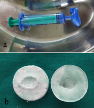
For this patient we decided to take impression using Light body Addition silicone material. ([Figure 2] a) For taking impression patient is asked to look straight and impression material injected into the selected impression using syringe and slowly fill the defect and patient is asked to move his eye slowly in all the directions .small amount of material should flow out from inner canthus. The impression was gently removed first by massaging the lower lid downwards and away from the nose first and then sliding the impression out from the upper eyelid in an arc like path. The impression was then washed and disinfected with Revita lens solution (Ocutec, UK).
Making a stone model
A stone model is made of the impression. Take the stone in small mold fill it an dthen place the impression over it and remove the impression from the syringe. Then apply vaseline over the forst pour and then second pour is poured over the first pour in small mold itself. ([Figure 2]b)
Making a wax pattern
Model which is made is used for wax pattern fabrication. a light yellow wax (Technovent Ltd, southwales and UK) was pour over the first mold and second layer of mold. ([Figure 3]a) on hardening the wax pattern, retrieve it gently and smoothened with the help of a carver and guaze. Try the wax pattern in patients eye and adjust accordingly. It should not look bulky and nor even under filled.([Figure 3]b) wax pattern should fit in eye properly and should not come out while moving eye in all directions.
Attaching the iris
In this case colour matched iris selected from acrylic stock eye , and iris matched with adjacent eye and cut from stock eye and placed over the wax pattern with normal gaze. Iris should coinside with the size of adjacent eye (range 11-13 mm with an average of 12mm). while placing the patient should lookmedial and downward at this stage. Various techniques has been described in past for iris positioning. For this case we used graph method to place the iris. Once the iris is positioned it should flasked in two part flask using dental plaster. Curing is to be done according to conventional techique. ([Figure 4] a,b)
Colour matching done using tooth coloured heat cured acrylic resin and using acrylic paints sclera should match with the adjacent eye.
As patient is pediatric patient, instructions given to the parents, how to insert & remove the porsthesis without hurting the patient. Its also important to take care about the hygiene of the prosthesis. All the instructions given to the patient’s parents. It has also been explained that prosthesis might need repolishing at some intervals.


Discussion
Prosthesis given to the patient not just improves esthetics but also prevent social embarrasment. As in this case patient is pediatric patient so its important to maintain the size of the ocular defect otherwise it leads to microopthalmia and its then very difficult to restore such defects. technique described here is simple and can be easily tried.
Conclusion
A prosthetic eye is a ray of hope for patients with an ocular defects to restore esthetics and prevents psychological trauma. An ocular eye is not just restoring ethetics but also restoring remaining structures of eye. But inefficient to restore visison.
Source of Funding
None.
Conflict of Interest
None.
References
- G T Raflo, W Tasman, E Jarger. Enucleation and evisceration. Duane’s Clinical Ophthalmology 1995. [Google Scholar]
- S B Patil, R Meshramkar, B H Naveen, N P Patil. Ocular prosthesis: a brief review and fabrication of an ocular prosthesis for a geriatric patient. Gerodontology 2008. [Google Scholar]
- S. Taicher, H.M. Steinberg, I. Tubiana, M. Sela. Modified stock-eye ocular prosthesis. J Prosth Dent 1985. [Google Scholar]
- S Lal, A Schwartz, T Gandhi, M Moss. Maxillofacial Prosthodontics for the Pediatric Patient:"An Eye-Opening Experience". J Clin Pediatr Dent 2007. [Google Scholar]
- S S Guttal, S M Joshi, L K Pillai, R K Nadiger, Gerodontology. Ocular prosthesis for a geriatric patient with customised iris: A report of two cases. Gerodontology 2011. [Google Scholar]
- A Ioli-Ioanna, P C Montgomery, P J Wesley, J C Lemon. Digital imaging in the fabrication of ocular prostheses. J Prosth Dent 2006. [Google Scholar]
- S O Bartlett, D J Moore. Ocular prosthesis: A physiologic system. J Prosth Dent 1973. [Google Scholar]
- K I Perman, H I Baylis. Evisceration, Enucleation, and Exenteration. Otolaryngologic Clin North Am 1988. [Google Scholar]
