- Visibility 3.1k Views
- Downloads 126 Downloads
- Permissions
- DOI 10.18231/j.idjsr.2021.033
-
CrossMark

Abstract
In routine orthodontics practice anchorage is a critical factors that determining the success of an orthodontic treatment. For many years’ anchorage has been a strenuous factor. Many modalities have been tried for preventing the anchorage loss by using the extra oral and intraoral devices according to various scientific literature, yet the orthodontists did not find any convenient solutions to solve this problem until the mini- implants were emerged to the speciality. During the early 20th century introduction of various skeletal anchorage devices which includes prosthetic implants, palatal implants, mini-plates and screws. The implants used in orthodontics for the purpose of effective treatment are also known as Temporary Anchorage Devices (TADs), which helps in bringing down the strenuous efforts employed to prevent anchorage loss and they are easy and small and can be insert and remove without stenos efforts, they can be immediately loaded after insertion, and they can provide absolute anchorage for orthodontic treatment, which require minimal need patient compliance there by increasing its popularity. This article gives a brief reviews of indications, contraindications, safety zones for TADs, their insertion procedure, complications, failures and medico legal aspects.
Introduction
For a success of orthodontic treatment anchorage is one of the main factors. Conservation of orthodontic anchorage has been a one of the perennial problem for orthodontist. Conventional techniques use either intra-oral sites or extra oral means. For optimum treatment results various approaches have been employed which includes using implants for anchorage with varying success. Extra oral anchorage is cumbersome to use and it usually requires patient’s compliance and may cause injury during their use. The term ‘Absolute anchorage’ can be defined as when the anchorage unit remains completely stable.
The skeletal Anchorage used in orthodontics are of absolute anchorage which is achieved with the use of orthodontic mini- implants. With the appropriate use of Orthodontic mini-implants, maximum anchorage is possible which will reduces the unwanted side-effects. [1]
Mini-screws are also known as TAD’S (Temporary Anchorage Device) or Micro-implants or Ortho-implant, by the advent of TADS there is a significant revolution in the field of clinical Orthodontics.
In 1945, Gainsforth & Higley conducted a study in which Vitallium screws & SS wires in the Ramal area of the dog’s mandible so as to bring about retraction of upper canines. This was considered to be the first published case where implants are used for orthodontic anchorage. [2] In 1984, Robert & fellow researchers collaborated with the findings of Branemark where they placed titanium implants in rabbits. The study concluded that titanium endosseous implants provides firm osseous anchorage.[3] In 1988, Vitallium implant were used by Creekmore for anchorage for the purpose of intruding upper anterior teeth. [4]
Classification of Orthodontic Mini Implant.[5], [6]
Orthodontic implant are alloplastic material devices which are surgically inserted into or onto jaw bone and it is classified as:
Based on the Location
Subperiosteal they lies over the bony ridge. Onplants are the subperiosteal design that are used for orthodontic purposes.
Transosseous; These implant body penetrates the mandible completely.
Endosseous/ Endosteal are those implants that are partially submerged and are anchored within bone.
Based on the form
Solid
Hollow
Vented
According to the composition
Stainless steel,
Cobalt-Chromium-Molybdenum,
Titanium,
Ceramic Implants.
Miscellaneous such as Vitreous carbon and composites
According to the surface structure
Threaded or Non-threaded
Porous or Non Porous.
5) Based on head type –
Small head type
Long head type
Circle head type
Fixation head type
Bracket head type
According to March 2005 classification –
Biocompatible TADS
Biological TADS
Parts of orthodontic implant
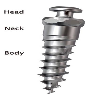
Orthodontic mini implant screw/plate has three parts
Implant head, this is the part where the implant is attached to the driver and the head of the implant serves as the abutment and could be the source of attachment for elastics/ coil-springs
Neck –It is the junction between head of the implant and platform for attachment of an elastic, NiTi coil spring or other accessories.
Body – It is parallel in shape and self- drilling with wide diameter and have deep thread pitches. It provides better anchorage, good mechanical retention, less loosening breakage. It is the part of mini implant which get embedded inside bone.
Ideal requirements for implant biomaterial
The following are the ideal requirements for implant biomaterial –
Biological properties
Physical properties
Biological properties –
Provide effective Osseo integration.
Shouldn’t cause any harm to soft tissue and hard tissue.
Should not contain the toxic diffusible substance.
Should be free agents that may cause an allergic reaction.
Should have no carcinogenic potential.
Should be tasteless and odourless.
Physical properties –
Should be dimensionally stable.
Should possess adequate strength and resilience
Should able to resist biting or chewing forces.
The osseointegrating orthodontic / dental implants / screws are composed of 99% titanium. The medical grade titanium used are of grade I to IV.
Commercially pure titanium (C P Ti) is used widely for implants fabrication because it possesses excellent biocompatibility and suitable mechanical properties. Use of Ti grades I to IV in for the manufacturing of non –osseointegrated / mechanical retentive miniscrews showns failures as screw were thinner.
Therefore, the titanium alloy (Ti - 6Al - 4V) (grade V) is the material for orthodontic miniscrews / mini implants. Titanium alloy (Ti - 6Al - 4V) increases the modulus of elasticity to six times that of bone so that long and thin
Indications for implant in orthodontics
Indications for implants in orthodontics are as follows
Retract and align anterior teeth
In first molar extraction sites they are used for closing the edentulous spaces
Intrude or extrude teeth,
Protract or retract teeth of one arch,
Stabilizing the teeth with less bone support,
Contraindication for implant placement
There is no absolute contraindication for orthodontic mini implant placement the placement of implant are contraindicated in cases of Psychiatric diseases (psychoses dysmorphobia severe systemic disorder like osteoporosis, blood disorders ,alcoholics ,drug abusers. Patients with poor bone quality and diabetic patients.
Treatment Considerations [6], [7], [8]
Age of the patient
The age of the patients is an important consideration for implant placement in growing children the use of implant in the anterior maxilla is contraindicated due to opened mid palatal suture Resorption in the posterior part of the maxilla resulting to the exposure of the implants due to growth changes..=
Periodontal status
Patients with satisfactory periodontal status with adequate amount of bone support and thick compact cortical bone are indicated or mini care should be taken to maintain good oral hygiene.
Systemic manifestations
One of the predisposed factor for delayed wound healing in case of diabetics are destructive habits like smoking. In case of chronic smokers it is contraindicated to placement the orthodontic mini-implant and it is noted that delayed or inadequate tissue healing and poor osseointegration is noted.
Radiographic analysis
During placement of orthodontic mini implants careful observation of Periapical pathology and Radiopaque/radiolucent should be examined and diagnosed in the regions above the inferior alveolar region, the maxillary sinus, adequate space above IAN or below maxillary sinus are to be taken care, During placement of mini-implant a minimum of 2mm from the inferior alveolar canal or below the maxillary sinus with adequate interradicular area should be there.
Safe zones for implant placement
The most commonly used placement sites for miniscrews in maxilla and mandible are as follows
In Maxilla: Inter radicular alveolar –as the buccal cortical bone on the entire maxillary alveolar process is about 3mm to 4mm, so longer screws are needed. Most commonly used sites are –
Between second premolar and first permanent molar
Between the first and second permanent molar
Between the two central incisors, used for intrusion
Infrazygomatic region – zygomatic buttress
Palatal areas.
Maxillary tuberosity region
Mid palatal area
In Mandible: In the mandible dense cortical bone on the buccal area is present, so the screws of smaller in size should be used, so the possibility of root contact is remote. Most common sites are –
Between second premolar and first permanent molar
Between first and second permanent molar
Between two central incisors
Between mandibular canine and premolar buccally
Retromolar area
Mandibular symphysis facially
Some of the anatomical and vital structures that should be kept care of during micro-implant placement includes- inferior alveolar nerve, artery, vein, mental foramen, maxillary sinus and nasal cavity. [9]
Implant placement angulation
In Maxilla: micro- implant is placed at an angulation of 30- degree to 40 -degree angle to the long axis of the teeth in the maxilla, it will keep the screw in the widest space available between the roots apically.
In the Mandible, Micro implants are placed at an angulation of 10- degree to 20- degree because the buccal cortex is of dense bone and curves out more buccally from gingival margins. So mini screws of shorter dimension can be used than those used in the maxilla. Also the angle is reduced to 10- degree to 20- degree with little risk of touching the roots.
Methods of placing micro screws / micro implants
The method of placement of orthodontic miniscrew into the alveolar bone depends upon the type of screw chosen. There are two different types of screws available –
Self-Tapping: First a tunnel is drilled into the bone with the help of pilot drill and then implant is driven into tunnel
Tips: blunt, smooth and rounded
Threads: - thick rounded and blunt
Self-Drilling: Here the implant itself acts as a drill and it is directly inserted into bone Tips: - sharp, hooked and pointed
Threads: - thin, pointed
Procedure for microimplant placement
The various steps for surgical implant placement are as follows
Topical Anaesthesia: Soft local Infiltration usually adequate
Aseptic preparation – A disinfecting agent can be used to prepare an intraoral or extra oral site for keeping the surgical area aseptic.
Drilling –Mini implants are loaded to the selected micro screw driver, and the screw is inserted at the desired location. Guide bar can be use and placed on the tooth before exposing the patient to IOPA. The guide is placed during micro implant insertion it should be retained, so it can help in placement of a micro- implant. The direction of insertion is first at 90 degrees to occlusal plane and then angulated at 30 - 40 degree in the maxilla and in case of mandible at 10- 20- degree. To ensure proper stability of implants wobbling in the axis of a driver should be avoided. During placement screw should be smooth alternating between turns and stops
Loading of implants
Two types of loading can be employed which includes immediate loading and delayed loading in terms of orthodontic mini-implants, the primary stability is more important than the Osseo integration. Clinical studies have shown that there is no significant difference exists between the immediate loading and delayed loading when the force levels are between 200- 300gms after achieving primary stability. However, it may be better to wait approximately 2-3 weeks for soft tissue healing.
Stability of orthodontic implants
In cases of orthodontic mini implant 2 types of stability are seen they are Primary stability and the secondary stability.
Primary stability or initial stability is noted immediately after the insertion of an orthodontic mini-implant. Which is the prime factor consideration for healing and loading. The factors that contributing and are responsible for achieving the primary stability includes- Implant diameter, the length of implant, and the number of flutes and design of threads, cortical bone thickness and also the bone density. Primary stability also depends on the placement technique and location of implant placement.[10], [11], [12], [13], [14]
Secondary stability is seen after implant placement and the bone regeneration and remodelling which contributes to increasing the stability. [15]
Complication of orthodontic implants
Complications can arise during the placement and after orthodontic loading of TAD’s in regard to stability and patient safety.
Complications during Insertion includes
Trauma to the dental root or to periodontal ligament
Orthodontic implant slippage
Nerve involvement
Air subcutaneous emphysema
Nasal and maxillary sinus perforation
Implants bending, fracture, and torsional stress
Complications under Orthodontic Loading
Miniscrew migration
Soft-Tissue Complications
Aphthous ulceration
Soft tissue inflammation, infection, and peri implantitis
There can be soft-tissue coverage on to the head of the mini implant and auxiliary
Complications During Removal
Screw fracture
Partial osseointegration
Conclusion
The introduction of orthodontic mini implants on the field of dentistry had a tremendous impact on dental treatment plans. Mini implants help the orthodontist to overcome the unwanted reciprocal tooth movement happening during routine dental treatment. The presently available implant systems are ease of placement (able to be placed by orthodontist), least invasive procedure, and best physical design properties to deliver optimum mechanical forces. bound to change and evolve into more patient friendly and operator convenient designs. Long-term clinical trials are awaited to establish clinical guidelines in using implants for both orthodontic and orthopaedic anchorage.
Conflicts of Interest
The authors declare that there are no conflicts of interest regarding the publication of this paper.
Source of Funding
None.
References
- Samuel R, Jones ML. Orthodontic facebow injuries and safety equipment. Eur J Orthod. 1994;16(5):385-94. [Google Scholar] [Crossref]
- Gainsforth BL, Highley LB. A study of orthodontic anchorage possibilities in basal bone. Am J Orthod. 1945;31:406-23. [Google Scholar] [Crossref]
- Roberts WE, Smith RK, Silberman Y, Mozsary PG, Smith RS. Osseous adaptation to continuous loading of rigid endosseous implants. Am J Orthodont. 1984;86(2):95-111. [Google Scholar] [Crossref]
- Creekmore TD, Eklund MK. The possibility of skeletal anchorage. J Clin Orthod. 1983;17(4):266-9. [Google Scholar]
- Wilmes B, Ottenstreuer S, Su YY, Drescher D. Impact of implant design on primary stability of orthodontic mini-implants. J Orofac Orthop. 2008;69(1):42-50. [Google Scholar] [Crossref]
- Melsen B, Costa A. Immediate loading of implants usedfor orthodontic anchorage. Clin Orthod Res. 2000;3(1):23-8. [Google Scholar] [Crossref]
- Wilmes B, BL, SB, SJB. Fields of application of mini-implants, in Mini- Implants in Orthodontics: Innovative Anchorage Concepts. . 2008. [Google Scholar]
- Kanomi R. Mini-implant for orthodontic anchorage. J Clin Orthod. 1997;31(11):763-7. [Google Scholar]
- Poggioa PM, Incorvati C, Velo S, Carano A. A Guide for Miniscrew Positioning in the Maxillary and Mandibular Arch. Angle Orthod. 2006;76(2):191-7. [Google Scholar] [Crossref]
- Wilmes B, Drescher D. Vertical periodontal ligament distraction-a new method for aligning ankylosed and displaced canines. J Orofac Orthop. 2009;70(3):213-23. [Google Scholar] [Crossref]
- Berens A, Wiechmann D, Dempf R. Mini- and microscrews for temporary skeletal anchorage in orthodontic therapy. J Orofac Orthop. 2006;67(6):450-8. [Google Scholar] [Crossref]
- Cheng SJ, Tseng IY, Lee JJ, Kok SH. A prospective study of the risk factors associated with failure of miniimplants used for orthodontic anchorage. Int J Oral Maxillofac Impl. 2004;19(1):100-6. [Google Scholar]
- Fritz U, Ehmer A, Diedrich P. Clinical suitability of titanium microscrews for orthodontic anchorage-preliminary experiences. J Orofac Orthop. 2004;65(5):410-8. [Google Scholar] [Crossref]
- Berens A, Wiechmann D, Rudiger J. Skeletal anchorage in orthodontics with mini and microscrews. Int Orthod. 2005;3(3):235-43. [Google Scholar]
- Wiechmann D, Meyer U, Buchter A. Success rate of mini- and micro-implants used for orthodontic anchorage: A prospective clinical study. Clin Oral Impl Res. 2007;18(2):263-7. [Google Scholar] [Crossref]
- Abstract
- Introduction
- Classification of Orthodontic Mini Implant.[5], [6]
- Parts of orthodontic implant
- Orthodontic mini implant screw/plate has three parts
- Ideal requirements for implant biomaterial
- Indications for implant in orthodontics
- Contraindication for implant placement
- Treatment Considerations [6], [7], [8]
- Age of the patient
- Periodontal status
- Systemic manifestations
- Radiographic analysis
- Safe zones for implant placement
- Implant placement angulation
- Loading of implants
- Stability of orthodontic implants
- Complication of orthodontic implants
- Conclusion
- Conflicts of Interest
- Source of Funding
- References